
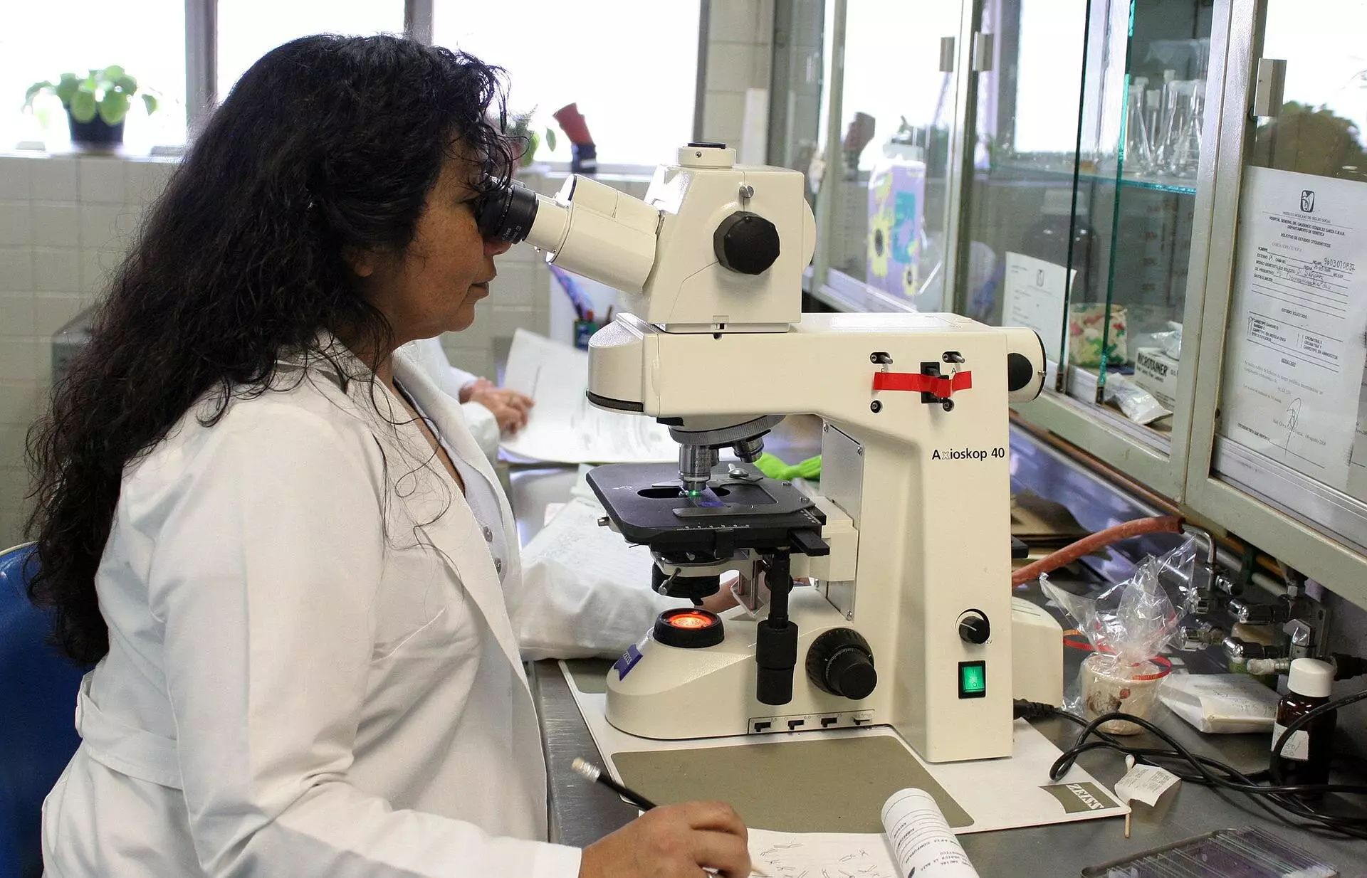
When using a microscope to view biological samples, the medium in which the lens of the objective is located can significantly impact the accuracy of the observations. If the lens is surrounded by a medium that is different from the sample being viewed, such as air instead of water, the light rays will bend differently. This can result in the measured depth of the sample appearing smaller than it actually is, causing the sample to look flattened.
For decades, theories have been developed to address this issue by determining a corrective factor for the depth measurements. However, these theories have all assumed that the corrective factor remains constant regardless of the sample’s depth. It wasn’t until Nobel laureate Stefan Hell pointed out in the 90s that this scaling could be depth-dependent that researchers began to reevaluate their assumptions.
Recent research conducted by Sergey Loginov, a former postdoc at Delft University of Technology, has shown through calculations and a mathematical model that the flattening effect is indeed more pronounced closer to the lens than farther away. Subsequent experiments conducted by Ph.D. candidate Daan Boltje and postdoc Ernest van der Wee in the lab confirmed that the corrective factor is depth-dependent. These findings have been published in the journal Optica.
The research team has developed a web tool and software that allows researchers to determine the precise corrective factor for their experiments. This tool has proven to be invaluable in cutting out proteins and their surroundings from biological systems for analysis with electron microscopy. By accurately determining the depth of samples, researchers can save time and resources by focusing only on relevant proteins and biological structures.
Understanding the precise structure of proteins in biological systems is crucial for gaining insights into abnormalities and diseases. By using depth-dependent scaling factors in microscopy, researchers can enhance their ability to study and combat these health issues. The ability to accurately view and analyze biological structures can lead to significant advancements in the field of biology and medicine.
The web tool developed by the research team allows users to input specific details about their experiments, such as refractive indices, aperture angles, and the wavelength of light used. The tool then generates a curve depicting the depth-dependent scaling factor, which can be exported for further analysis. Researchers can also compare the results with existing theories to gain a comprehensive understanding of the issue.
The research on depth-dependent scaling in microscopy represents a significant advancement in the field. By recognizing and addressing the limitations of traditional theories, researchers can improve the accuracy and efficiency of their experiments. The development of practical tools like the web tool created by the research team demonstrates the importance of collaboration between theory and experimentation in driving scientific progress.
Leave a Reply