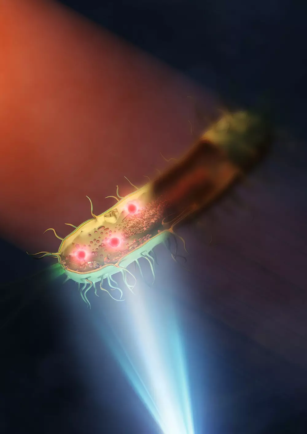
Microscopy has long been a crucial tool in the field of biological research, allowing scientists to explore the intricate structures inside living organisms. However, traditional microscopy techniques have often been limited by their resolution capabilities. One such technique, mid-infrared microscopy, has faced challenges in providing high-resolution images of live cells. Despite its potential to offer both chemical and structural information without the need for labeling or damaging samples, mid-infrared microscopy has lagged behind other more advanced microscopy methods.
Super-resolution fluorescent microscopes, for example, require samples to be labeled with fluorescence, which can be harmful to the specimen. Additionally, prolonged exposure to light during imaging can lead to sample bleaching, rendering them unusable for further study. On the other hand, electron microscopes offer impressive detail but require samples to be placed in a vacuum, prohibiting the study of live cells. These limitations have underscored the need for the development of improved microscopy techniques that can provide high-resolution images of live cells without damaging them.
A team of researchers at the University of Tokyo has made a significant breakthrough in the field of mid-infrared microscopy. By utilizing a synthetic aperture technique, the team was able to improve the resolution of mid-infrared microscopy by up to 30-fold, achieving a spatial resolution of 120 nanometers. This advancement is a major leap forward in the field of microscopy, allowing scientists to observe the intracellular structures of living bacteria with unprecedented clarity.
Implications for Research
The enhanced resolution capabilities of this new mid-infrared microscope hold great promise for a wide range of research fields. The ability to visualize samples at such a small scale can aid in the study of infectious diseases, antimicrobial resistance, and other crucial areas of research. Furthermore, this breakthrough paves the way for the development of even more accurate mid-infrared-based imaging techniques in the future.
Professor Takuro Ideguchi, one of the researchers involved in the project, expressed optimism about the future of mid-infrared microscopy. By using better lenses and shorter wavelengths of visible light, the spatial resolution of the microscope could potentially be improved to below 100 nanometers. This opens up exciting possibilities for further advancements in the field of microscopy and has the potential to revolutionize our understanding of the microscopic world.
The development of an improved mid-infrared microscope by researchers at the University of Tokyo represents a significant milestone in the field of microscopy. By overcoming the limitations of traditional mid-infrared microscopy, this breakthrough has the potential to greatly impact various areas of scientific research. The enhanced resolution capabilities of this new microscope offer a unique opportunity to explore the intricate structures inside living cells with unprecedented clarity. As further advancements are made in the field of microscopy, our ability to study and understand the microscopic world will continue to expand, opening up new possibilities for scientific discovery and innovation.


Leave a Reply