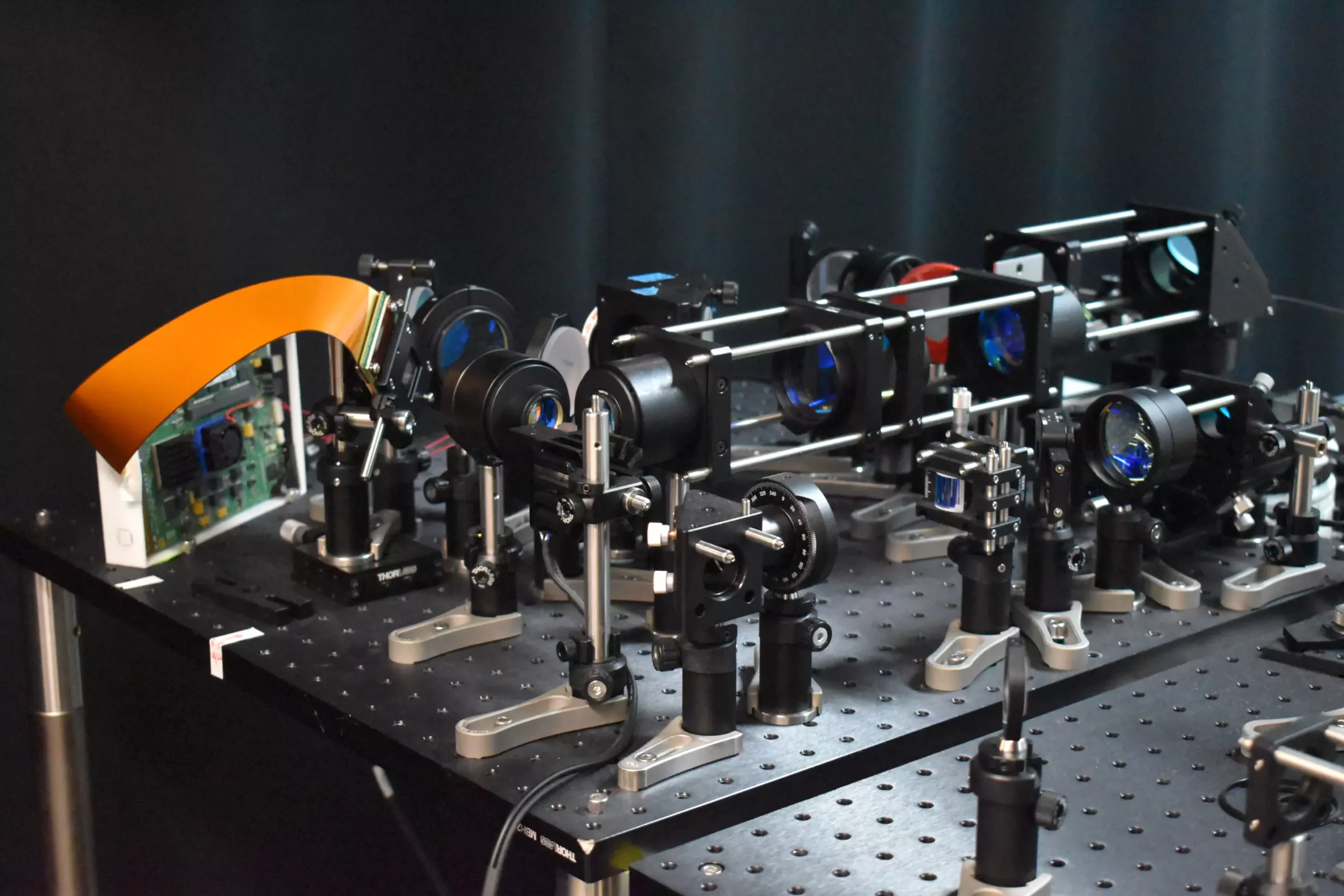
Neuroimaging has taken a significant leap forward with the development of a new two-photon fluorescence microscope. This groundbreaking technology has the capability to capture high-speed images of neural activity at cellular resolution, providing researchers with a clearer and more detailed view of how neurons communicate in real-time.
Advantages Over Traditional Microscopy Techniques
Unlike traditional two-photon microscopy, this new approach allows for imaging at much faster speeds and with significantly less harm to brain tissue. By incorporating a new adaptive sampling scheme and replacing point illumination with line illumination, researchers have been able to image neuronal activity in a mouse cortex in vivo at speeds ten times faster than before, while reducing the laser power on the brain more than tenfold.
The implications of this new technology are vast. It has the potential to enhance our understanding of fundamental brain functions such as learning, memory, and decision-making. By observing neural activity in real-time, researchers can gain valuable insights into the communication and interaction among different neurons during these processes. Additionally, this technology could be instrumental in studying the pathology of neurological diseases at their earliest stages, leading to more effective treatments for conditions such as Alzheimer’s, Parkinson’s, and epilepsy.
The Key Innovation: Adaptive Sampling
One of the key innovations of this new microscope is the adaptive sampling strategy. Rather than using a point of light to scan the brain, researchers now use a short line of light to illuminate specific areas where neurons are active, enabling a larger area to be imaged at once. This not only speeds up the imaging process but also reduces the total light energy deposited into the brain tissue, minimizing the risk of damage. By using a digital micromirror device to shape and steer the light beam, researchers can precisely target active neurons and achieve adaptive sampling by adjusting to the neuronal structure of the brain tissue being imaged.
By combining the adaptive line-excitation technique with advanced computational algorithms, researchers are able to isolate the activity of individual neurons, providing a more accurate interpretation of complex neural interactions and the functional architecture of the brain. This new microscope has the capability to capture calcium signals, indicators of neural activity, at a speed of 198 Hz, significantly faster than traditional methods. This speed allows for the monitoring of rapid neuronal events that may be missed by slower imaging techniques.
Looking ahead, the researchers are working on integrating voltage imaging capabilities into the microscope to capture rapid readouts of neural activity. They plan to use this technology for real neuroscience applications, such as observing neural activity during learning and studying brain activity in disease states. Furthermore, efforts are underway to improve the user-friendliness and reduce the size of the microscope to enhance its utility in neuroscience research.
The development of this new two-photon fluorescence microscope represents a significant advancement in the field of neuroimaging. With its ability to capture high-speed images of neural activity at cellular resolution, this technology has the potential to revolutionize our understanding of brain function and neurological diseases. As researchers continue to explore its applications and push the boundaries of what is possible, the future of neuroscience looks brighter than ever before.


Leave a Reply