
In a groundbreaking study published in the journal Optica, researchers at HHMI’s Janelia Research Campus have introduced a new approach to microscopy by applying techniques commonly used in astronomy to unblur images of far-away galaxies. This innovative adaptation aims to provide biologists with a faster and more cost-effective method to obtain clearer and sharper microscopy images, revolutionizing the field of life sciences.
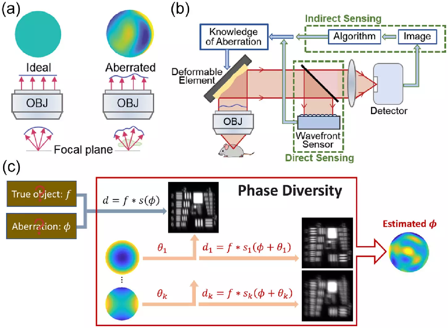
Astronomers have long utilized techniques to enhance the clarity and sharpness of images captured by telescopes of distant galaxies. By measuring how light is distorted by the atmosphere and applying corrections to eliminate aberrations, astronomers can produce high-resolution images. In the realm of microscopy, researchers have been incorporating similar adaptive optics methods to enhance images of thick biological samples that also distort light. However, these techniques, known as adaptive optics, have been hindered by their complexity, high cost, and slow processing speed, making them inaccessible to many research laboratories.
In an effort to democratize adaptive optics in biology, the team at HHMI’s Janelia Research Campus has turned to a different class of techniques known as phase diversity. Although phase diversity has been widely utilized in astronomy, it is relatively new in the life sciences. These methods involve adding additional images with known aberrations to a blurry image with unknown aberrations, providing valuable additional information to deblur the original image. Unlike conventional adaptive optics approaches, phase diversity does not require major modifications to the imaging system, making it a promising avenue for microscopy advancement.
To implement the new method, the research team first adapted the astronomy algorithm for microscopy applications and validated it through simulations. Subsequently, they developed a microscope equipped with a deformable mirror and two additional lenses to introduce known aberrations. These minor modifications to the existing microscope setup facilitate the phase diversity correction process. Additionally, the team enhanced the software used to execute the correction, ensuring optimal performance.
In their validation experiments, the researchers demonstrated that the new method significantly accelerated the calibration of the microscope’s deformable mirror compared to existing techniques. Furthermore, they successfully corrected randomly generated aberrations, resulting in clearer images of fluorescent beads and fixed cells. The next phase of research will involve testing the method on real-world biological samples, including living cells and tissues, and expanding its application to more sophisticated microscopy setups. The team also aims to streamline the method, making it more automated and user-friendly.
By offering a faster and more cost-effective alternative to current adaptive optics techniques, the novel method developed by the team at Janelia Research Campus has the potential to democratize the use of adaptive optics in biology. By providing researchers with clearer and more precise imaging capabilities, this innovation promises to revolutionize the field of microscopy, enabling biologists to explore and understand complex biological structures with unprecedented clarity and depth. Ultimately, this advancement in adaptive optics technology will drive forward scientific discovery and pave the way for new breakthroughs in the life sciences.
Leave a Reply